
核子醫學暨分子影像雜誌/Annals of Nuclear Medicine and Molecular Imaging
中華民國核醫學學會 & Ainosco Press,正常發行
選擇卷期
- 期刊
Remarkable progress in clinical applications of nuclear medicine theranostics has prompted a significant increase in global demand for positron emission tomography (PET) radiopharmaceuticals (drugs), such as the USA. Washington University School of Medicine has received a 5 years' project with US $ 6.3 million of financial support from National Institutes of Health since 2018 and establishes the PET-Radiotracer Translation & Resource Center (PET-RTRC) to provide assistance in the development and clinical trials of new PET drugs in the USA. Additionally, they hold related workshops to meet the objects of PET-RTRC. The workshops were only opened to domestic researchers in the past; however, it was opened to foreign participants in 2020. Because the USA and Europe policies for development and clinical use of PET drugs are always important references for Taiwan researchers working in nuclear medicine fields, we attended the PET-RTRC workshop this year and collected the latest information, which includes current US regulations/bylaws in compounding and clinical trials of PET new drugs, the procedures of documental applications and facility requirements of on-site preparation of PET drugs, as well as the differences between current regulatory situations of Taiwan and the USA.
- 期刊
Background: Radiotherapy (RT) and chemotherapy can improve tumor control for breast cancer (BC). Whether it is chemotherapy or RT, they may increase the incidence of cardiotoxicity and may cause complications, such as cardiovascular disease, myocardial perfusion defect, or life-threatening heart failure. Previous studies demonstrated that single-photon emission computed tomography myocardial perfusion imaging (SPECT MPI) can early detect myocardial dysfunction caused after treatment. Therefore, in order to comprehend the cardiac function of before RT for BC patients, this study aims to retrospectively collect cardiac function indexes of BC patients before RT and analyze the difference between with and without chemotherapy patients. Methods: This retrospective study included 49 left BC patients who received SPECT MPI before RT, and record wall motion (WM), wall thickening (WT), motion scores, and thickening scores. The cardiac function indexes between with and without chemotherapy were analyzed. Results: Preliminary results showed that 32 patients were with chemotherapy and 17 patients were without chemotherapy. The WM was 8.21 ± 2.30 mm and 8.55 ± 2.21 mm (p = 0.502) respectively; the WT was 50.58 ± 21.04% and 54.51 ± 20.82% (p = 0.488). Besides, there was WM abnormality (motion scores ≥ 2) with chemotherapy patients in segments 2, 3, 4, 5, 6, 8, 9, and 12 and without chemotherapy patients in segments 2, 3, 4, and 8. After chemotherapy, patients with thickening abnormalities (thickening scores ≥ 2) were in segments 2, 3, 4, 5, 6, and 8; patients without chemotherapy were in segment 3. Conclusions: This retrospective study found that patients with chemotherapy were reducing the cardiac function. Therefore, when the patients receive the following RT, the radiation dose of the heart needs to be noticed because of chemotherapy.
- 期刊
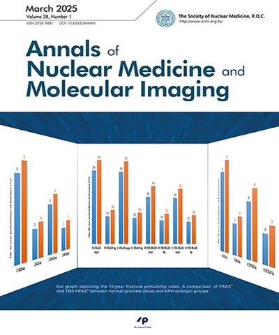
Background: In clinical practice for the bone mineral density (BMD) evaluation, lumbar spine and bilateral hip areas, were included. However, the fracture risk assessment tool (FRAX) only adapts the hip areas as calculation factor. That is, for cases with osteoporosis in lumbar spine but normal in hip areas, FRAX might underestimate the risk of spinal fracture. The aim of this study analyzes the difference between low fracture risk (major osteoporotic fracture risk < 20%, and hip fracture risk < 3%) and high-risk group according to FRAX in osteoporotic patients. Methods: This retrospective research study, outpatient records and dual-energy X-ray absorptiometry (DXA) with variable for FRAX (age, gender, height, weight, previous fracture, smoking, glucocorticoids, rheumatoid arthritis, alcohol and high fracture probabilities) from November 2014 to December 2019 were collected. Past history of spinal surgery and total hip replacement or incomplete medical records were excluded. Seven thousand one hundred forty-eight cases, 1,183 (17%) males and 5,965 (83%) females, were included for analysis. Results: There showed older, higher, heavier, higher cigarette and alcohol taking rate, lower rheumatoid arthritis rate and lower fracture probabilities in men than in women with significant difference (p < 0.05). Significant difference in the BMD and FRAX fracture probabilities between normal, low bone density, and osteoporosis groups were also noted. In osteoporotic cases, older, shorter, thinner, less BMD/T-score of bilateral femoral neck and bilateral total femurs in high fracture risk group than low fracture risk group with significant difference (p < 0.05). Four point four percent patients with low FRAX risk were diagnosed to be osteoporosis solely because of the lumbar BMD measurement was no more than -2.5. In addition, the demographic variables in the osteoporotic patients showed significant difference between high FRAX risk group and low FRAX risk group. Conclusions: In this study, the osteoporotic cases defined by lumbar vertebrae DXA results with fair bilateral hip DXA results are classified into low fracture risk group. Therefore, three areas (lumbar vertebrae and bilateral hips) DXA scan and 10-years fracture risk calculation for bilateral hips should be performed together to provide clinicians with accurate reference for patient treatment and medication.
- 期刊
Most neuroendocrine tumors (NETs) express somatostatin receptors and can be visualized by Gallium- 68-labeled somatostatin analogs positron emission tomography-computed tomography (PET/CT). We reported a case of metastatic liver NETs receiving Ga-68-DOTATOCPET/ CT, which identified the primary focus. A 34-year-old female suffered from intermittent abdominal pain and liver lesions were found by abdominal sonography. Subsequent CT and magnetic resonance imaging (MRI) revealed hypervascular liver masses and metastatic NETs were confirmed by echo-guided liver biopsy. F-18 fluorodeoxyglucose (FDG)-PET/CT did not find any FDG avid lesions. The patient underwent radiofrequency ablation for the liver lesions, and received long-acting octreotide (20 mg intramuscular) treatment. Ga-68-DOTATOC-PET/CT was then performed and demonstrated the primary focus of the neuroendocrine tumor in the small intestine. Segmental resection of ileum was performed and confirmed the presence of the neuroendocrine tumor, G2. After surgery, the patient's abdominal pain subsided, but kept long-acting octreotide treatment.
- 期刊
F-18 FDG PET is a useful functional image technique to evaluate the disease extent in many cancers by detecting hypermetabolic lesions. In combination with anatomical imaging tools such as computed tomography (CT) or magnetic resonance image, extra anatomical information could be provided for better lesion localization. However, the relatively lower spatial resolution of the "attenuation correction" CT or respiratory motion artifacts in positron emission tomography (PET)/CT fusion imaging may sometimes fail to correctly portray the real anatomical relationship between the lesion and nearby structures. We presented a case, in which the "attenuation correction" CT incorrectly delineated the anatomical relationship, misleading to an inaccurate impression. We recommend PET with contrast-enhanced CT, with focal full diagnostic CT, or with respiratory gating techniques might be an option in selected cases to avoid this kind of pitfall.
- 期刊
Here we describe a case of a 19-year-old young man with acute pulmonary embolism and left lower leg thrombosis after lower limb trauma post thrombolytic therapy and persistent D-Dimer elevation. Radionuclide venography revealed deep venous thrombosis in the left infra-popliteal to iliofemoral veins with good collaterals. Lower extremity ultrasound showed thrombosis over the left iliofemoral vein and downstream veins. We found that the left greater saphenous vein was an alternative pathway of collateral systems in the presence of deep vein thrombosis at the infra-popliteal vein.
- 期刊
A 41-year-old male with congenital osteosclerosis (CO) received open reduction and internal fixation for a right femur fracture then complicated with wound infection. Three years previously, he received the Girdlestone procedure but had discharging sinus ever since. Wound culture showed Staphylococcus aureus and Peptostreptococcus prevotii. But infection persisted even after vancomycin and teicoplanin treatment. Bone scan with three-phase study and single photon emission tomography/computed tomography (SPECT/CT) showed CO with osteomyelitis (OM) in the right proximal femur. Magnetic resonance imaging confirmed osteomyelitis. Adding a dynamic phase can differentiate OM and early skeletal uptake in CO. SPECT/CT can also help verify OM better than conventional imaging.