
核子醫學暨分子影像雜誌/Annals of Nuclear Medicine and Molecular Imaging
中華民國核醫學學會 & Ainosco Press,正常發行
選擇卷期
- 期刊
Positron emission tomography (PET) drugs play increasingly more roles in the application of modern medical treatments. Therefore, regulatory officers are implementing increased controls over PET drug productions to ensure patients' safety and announcing more regulatory requirements for PET drug manufacturers to follow. It is important for PET drug manufacturers to be compliant with current good manufacturing practice (cGMP). However, there are limited resources, so how to prioritize resources on the needed areas in response to this increasing regulatory burden has been widely discussed. Quality risk management (QRM) is a quality tool designed to use a risk- and scientific-based approach to systematically filter out higher priority items. Here, we introduce the fundamental structure of QRM and show some examples to demonstrate how to execute a QRM project. In addition, we provide some high-level concepts on how to integrate QRM into existing quality systems from a drug lifecycle perspective. Based on this article, PET drug manufacturers can apply QRM to efficiently control product quality and maintain compliance with cGMP requirements to ensure the safety of patients.
- 期刊
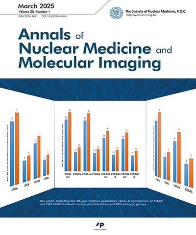
Background: According to the previous studies, we have separately explored the effects of benign prostate hypertrophy (BPH) and prostate specific antigens (PSA) on bone mineral density (BMD). Therefore, this study will further analyze the combination of BPH and PSA, and divide subjects into. BPH with PSA greater than 4 ng/mL (disease group) and no BPH with normal PSA (less than 4 ng/mL) (normal group). We compare the two variables at the same time to find whether the factors have a comprehensive impact on BMD. Methods: Total of 5,605 males who were examined in the preventive medical center of our hospital were collected for evaluation. Total hip replacement surgery, internal fixation in the lumbar spine or hip, lumbar vertebroplasty, or incomplete data were excluded. In the study, clinicians used ultrasound to confirm the diagnosis of BPH. The serum by chemiluminescence immunoassay is also used to determine whether the value of the PSA level is normal or not. PSA level greater than 4 ng/mL is considered abnormal. A dual-energy X-ray absorptiometer was used to measure the BMD of three parts (lumbar spine and bilateral hips). Results: Of the 5,605 males, 5,323 (95%) were in the normal group and 282 (5%) in the disease group. The mean age of patients was 53.85 ± 12.10 years in the normal group and 68.46 ± 7.72 years in the disease group (p < 0.001), with a significant difference. The diseased group having significantly increased BMD at the lumbar spine area, but significantly decreased BMD in the bilateral hips, as compared with the normal group. Conclusions: In conclusion, the disease group had high lumbar spine BMD but lower BMD in the bilateral hips. These findings suggest that men diagnosed with BPH or high PSA should be considered at risk for hip fractures and need the evaluation for BMD.
- 期刊
為落實全人照護,近年來臺灣各大醫院莫不持續深化跨領域團隊合作教育(interprofessional education, IPE)。在臨床實務上,分化良好型甲狀腺癌(well-differentiated thyroid cancer, WDTC)的治療除手術切除外,口服碘-131(radioiodine-131)已被證實為有效的輔助治療,其中碘-131治療尤其需要跨領域團隊合作方能竟全功。本文透過一次碘-131治療WDTC個案討論的IPE教學活動,闡述核醫科如何以跨領域團隊合作的做法處理一個臨床常見的問題,並在IPE納入團隊資源管理(team resource management)的精神,營造師生共學的教學環境。同時,我們也分析學員對此教學活動的認知和評估學員的學習成果,作為教學醫院推動IPE教學活動的參考,期望能透過此做法,增進相關同仁對團隊合作的認識,進而提升未來對甲狀腺癌病人的照護品質。
- 期刊
Splenosis is autotransplantation of splenic tissue after traumatic splenic rupture or splenectomy. It is often misdiagnosed as a tumor on imaging studies. Tc-99m heat-damaged red blood cells (RBCs) scintigraphy is a method with high specificity for functional splenic tissue. Using a single-photon-emission computed tomography (SPECT/ CT) technique, splenosis can be accurately confirmed to avoid invasive biopsy, resection, or treatment. A 71-year-old male patient is a case of hepatitis B and C virus-related liver disease. The patient has a history of splenectomy after blunt abdominal trauma. Liver tumors were found using sonography, and the patient’s alpha-fetoprotein (AFP) level was rising to 105 ng/mL. CT and magnetic resonance imaging revealed tumors in the S6 and the S2 of the liver and the left subphrenic region. After treatment of the S6 tumor, the patient’s AFP level returned to the normal range. Tc-99m heat-damaged RBCs SPECT/CT demonstrated that the S2 and the left subphrenic tumors were splenosis. A 60-year-old male patient was diagnosed with hepatitis C. The patient has a history of splenectomy after blunt abdominal trauma. Abdominal CT showed bilateral adrenal masses with peritoneal nodules. The initial impression was malignancy with peritoneal carcinomatosis. Because of the patient’s history of splenectomy, Tc-99m heat-damaged RBCs scintigraphy was suggested to rule out splenosis. After carefully identifying all adrenal and peritoneal lesions using SPECT/CT, the diagnosis of splenosis was confirmed. Tc-99m heat-damaged RBC scintigraphy with SPECT/CT can noninvasively confirm the diagnosis of splenosis to avoid invasive biopsy, resection, or treatment.
- 期刊
A 70-year-old smoking male presented with cough and hoarseness was diagnosed with adenocarcinoma of the left upper lobe of the lung. Fluorine-18 fluorodeoxyglucose (FDG) positron emission tomography/computed tomography (PET/CT) was performed for staging. In addition to FDG-avid primary lung tumor and left hilar lymph node, a focus of intense FDG uptake was incidentally noted at the posterior aspect of the left vocal cord. Laryngoscopy was done to exclude synchronous laryngeal malignancy. Normal appearance of the left vocal cord with contralateral vocal cord paralysis was found. Follow-up FDG PET/CT performed 2.5 years later revealed no discernible abnormal FDG uptake at the vocal cords, supported that the previously noted focal FDG uptake at the left vocal cord might be a benign etiology (e.g., compensatory overuse of the non-paralyzed vocal cord). One of the major causes of asymmetrical vocal cord FDG uptake is compensatory overuse of the unaffected vocal cord caused by unilateral vocal cord paralysis, which is not uncommon in patients with primary lung cancer and mediastinal surgery. We recommended nuclear physicians to be aware of this pitfall to avert a false-positive PET interpretation. Furthermore, diazepam administration or minimized vocalization before PET scan might be helpful to avoid this pitfall.