
核子醫學暨分子影像雜誌/Annals of Nuclear Medicine and Molecular Imaging
中華民國核醫學學會 & Ainosco Press,正常發行
選擇卷期
- 期刊
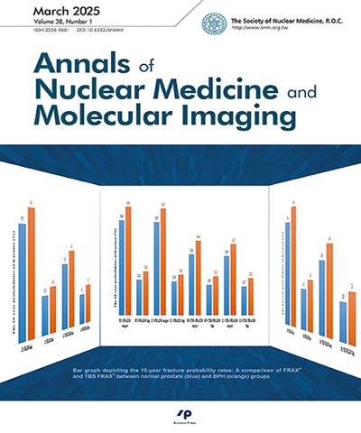
背景:本篇研究目的將探討三個部位(腰椎與雙側髖部骨質密度)均能分析與單一部位不能分析(腰椎與雙側髖部骨質密度任一部位)-因手術而無法分析,二組間各部位的骨質密度差異性,也進一步探討十年骨折率的變化。方法:本篇為回溯性研究,收集自2014年11月至2021年3月間,於臺灣南部某區域教學醫院,接受門診骨質密度檢測之受檢者,將受檢者分成二個組別,控制組(腰椎、左側髖部、右側髖部)三個部位均能分析,手術組(腰椎、左側髖部、右側髖部)只要一個部位開刀,不包含同時二個部位開刀,我們依據受檢者開刀部位分成二組分析。手術包括脊椎手術史(鋼釘內固定、椎體成形術或金屬植入物),髖部手術史(全髖關節置換手術或鋼釘內固定)。結果:本篇研究初步發現,利用骨質密度檢測三個部位,當有一部位因手術後無法評估時,與三個部位都能評估相比,將因手術部位影響骨密度評估,手術患者仍較控制組其骨質密度數值較低,骨折風險增加。結論:因此,本篇研究結論,研究發現金屬植入物固定手術後骨質密度會相較於控制組低,相對骨折率也比控制組高。因此未來建議臨床醫師定期追蹤金屬植入物術後骨質密度之變化,以降低再次骨折之狀況。
- 期刊
Background: The purpose of this research was to determine the cut-off value for head and neck (H/N) cancer by using semi-quantitative analysis and visual interpretation of skull base invasion. Methods: This is a retrospective study. From January 2011 to December 2012, we collected 1,015 consecutive patients referred for a whole body bone scan (WBS) to detect H/N cancer. Among them, 90 patients were female and 925 were male. We enrolled patients who received WBS and an additional single-photon emission computed tomography/computed tomography (SPECT/CT) of the H/ N area. Receiver operating characteristic (ROC) analysis of the area under the curve (AUC) was done to determine significance. Results: About 47.8% of planar bones showed visually abnormal uptake at the skull base on WBS. The remaining planar bone scans (52.2%) had normal findings at the skull base. Only about 26.1% of SPECT/CT showed visually abnormal uptake at the skull base, with 73.9% of normal findings at the skull base. ROC curve analysis showed the AUC of WBS was 62.6%, and that of the SPECT/CT was 89% (p < 0.005). The cut-off value was determined by ROC curve analysis. With a cut-off value of 1.965, the sensitivity of WBS was 73.6%, and the specificity was 44.9%. With a cut-off value of 6.94, the sensitivity of H/N SPECT/CT was 84.2%, and the specificity was 77.1%. Conclusions: Using semi-quantitative H/N SPECT/CT can facilitate the determination of the cut-off value for the interpretation of skull base invasion. This method was more sensitive than conventional bone scintigraphy and can help improve the confidence of nuclear medicine physicians in diagnosing H/N cancer. For patients suspected of having H/N cancer, more information can be obtained when a H/ N SPECT/CT is routinely added to WBS.
- 期刊
Peritoneal cerebrospinal fluid (CSF) pseudocyst is a complication of a ventriculoperitoneal shunt (VPS), which rarely occurs in adults, so such case reports are rare. We report here that the recurrence of hydrocephalus in an adult patient was caused by a rare peritoneal CSF pseudocyst, which was confirmed by subsequent scintigraphy and computed tomography (CT). A 60-year-old woman presented with clinically progressive weakness of the lower limbs, unable to walk, silence and slow speech, bilateral hearing impairment, and urinary incontinence, but no headache, diplopia, nausea, and vomiting. The results of brain CT showed hydrocephalus. Therefore, she underwent surgery to insert the VPS, and then she recovered as usual. One month later, she underwent a brain CT follow-up examination, which showed that the ventricles had shrunk. About 8 months after the shunt procedure, her previous symptoms reappeared. In the beginning, she also had fever, nausea, and vomiting. During the physical examination, only the muscle power of the limbs decreased (grade 3), and other neurological examinations were normal. The abdomen was oval in shape, without focal tenderness. When she was in the emergency room, her fever had subsided. The test results of blood and CSF were within the normal range. Subsequent CT of the brain confirmed the recurrence of hydrocephalus, so she was referred to the Department of Nuclear Medicine for VPS examination. A rare peritoneal CSF pseudocyst was accidentally discovered, which was confirmed by abdominal CT. After the patient received a new replacement of the VPS, the symptoms disappeared and the original function was restored.
- 期刊
A 34-year-old woman hospital personnel received her yearly follow-up fluorodeoxyglucose (FDG) positron emission tomography (PET) 4 days after the AstraZeneca COVID-19 vaccination. The PET images demonstrated new FDG uptakes at the left axilla and the left upper arm. After reviewing her clinical history, the FDG uptake at the left axillary lymph nodes (LNs) was attributed to the reaction to vaccination. This instance suggests that newly ipsilateral axillary LNs with FDG uptake after vaccination should be interpreted with caution. Vaccination history and the injection site should be inquired to avoid false-positive PET image interpretation and unwarranted biopsies.